Muscular System Front 24p Image Quiz. They cleanse the blood of toxins and balance the constituents of the circulation to homeostatic set points through the processes of filtration reabsorption and secretion.
Solved Assignment 10 Chapter 24 Art Labeling Activity Chegg Com
Anatomy of the Heart Ch.
. Youll notice familiar structures like the bladder and ureters as well as perhaps less familiar structures such as the renal artery and vein. Regulation of RBC production Activation of vitamin D. Figure 2521a Structure of the male urinary bladder and urethra.
The kidney diagram includes 5 versions one includes blood supply without any labels one without blood. The medullary pyramids contain collecting tubules ducts that travel towards the renal cortex carrying urine to exit the kidney. The blood supply to all these structures occurs through the branches and sub-branches of the renal artery called interlobular arteries and arcuate arteries respectively.
Blood enters the kidney via the paired renal arteries that form directly from the descending aorta and each enters the kidney at the renal hila. Figure 141 - ANS In the Nervous System See Diagram 2. Measuring renal clearance is one way in which clinicians can test renal function and qualify the.
This BUNDLE includes an oversized kidney diagram that is useful for reinforcement enrichment remediation for the most common kidney structures student notes a Review Chart as well as my Kidney Sketch Notes. Take a look at the urinary system diagram labeled below. Help Reset Proximal convoluted tubule Cortical nephron Renal corpuscle Juxtamedullary nephron Distal convoluted tubule Connecting tubules Nephron loop Collecting duct Papillary duct Renal papilla Urinary System Extracredit.
Anatomy and Physiology questions and answers. Overview image showing all of the main structures of the. The function of the digestive system is to break down the foods you eat release their nutrients and absorb those nutrients into the body.
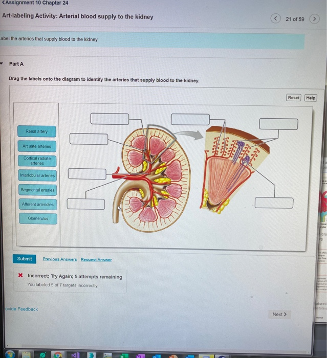
Reset Help Renal artery Arcuate arteries SA Cortical radiate Interlobular arteries. Figure 147 - Label the figure 3. Examining the Microscopic Anatomy of the Kidney Ureter and Urinary Bladder.
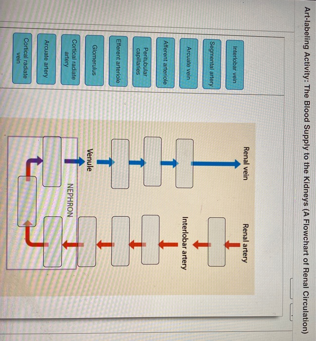
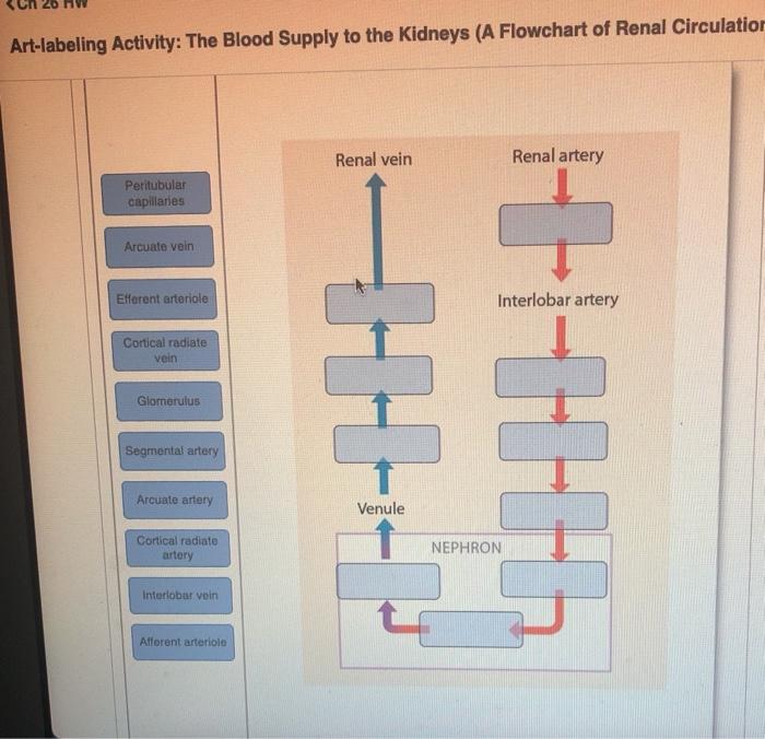
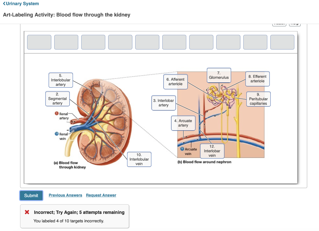
The IVC is formed by merging of the left and right common iliac veins at the L5 vertebral level just in front of the aortic bifurcation. Once in the kidney each renal artery first divides into segmental arteries followed by further branching to form interlobar arteries that pass through the renal columns to reach the cortex Figure 2513. Which one of the following is the correct gas exchange.
Identify the functional area of the kidney at letter B. Arteries of Cat Quiz By whitecollar Sporcle. Figure 2521b Structure of the female urinary bladder and urethra.
Nephrons are the functional units of the kidney. A number of other smaller veins empty into the left renal vein. Dissecting a Mammalian Kidney.
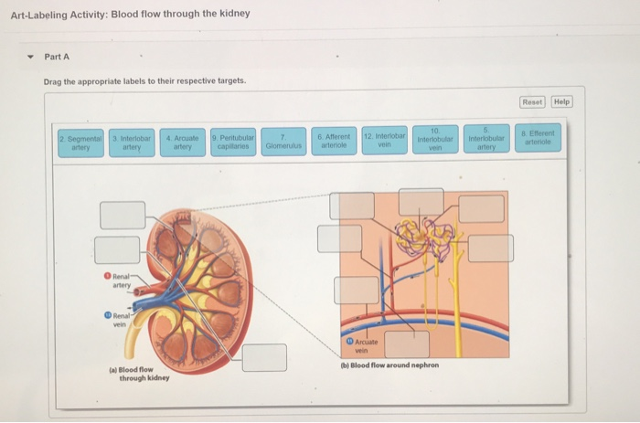
Blood supply to the kidneys. Performing a Hematocrit. The interlobular arteries supply blood to the borders of the cortex and medulla whereas the arcuate arteries diverge to form afferent arterioles that carry blood to the nephrons for filtration.
Where does the kidney filter the blood. Splanchnic circulation involves the blood supply that feeds and drains. Art Labeling Quiz Art Labeling Quiz This activity contains 7 questions.
12 Cranial Nerves Names In Order 12p Image Quiz. Renal hilum Renal pelvis. Exploring the Formed Elements of Blood.
Chapter 19 110 Essentials Figure. Blood flow through the kidney. C A micrograph shows the relative differences in thickness.
Blood supply from the kidneys flows into each renal vein normally the largest veins entering the inferior vena cava. It collects all the blood from the abdomen pelvis and lower limbs and carries it to the right atrium of the heart. The nephrons also function to control blood pressure via production of renin red blood cell production via the hormone erythropoetin and calcium absorption.
Although the small intestine is the workhorse of the system where the majority of digestion occurs and where most of the released nutrients are absorbed into the blood or lymph each of the digestive system organs makes a vital. Collects newly formed urine. Each adrenal vein drains the adrenal or suprarenal glands located immediately superior to the kidneys.
The inferior vena cava is the headmaster of the veins department. 12 Cranial Nerves Function 12p Image Quiz. This is due to its rich blood supplyit houses 9095 of the kidneys blood vessels.
Renal Clearance Proper kidney function is extremely important to maintaining homeostasis because the kidneys are responsible for eliminating metabolic wastes from the body and regulating fluid and electrolyte balance. The neurovascular and lymphatic supply of the peritoneum course to and from the posterior abdominal wall and gut tube through the two-layered mesentery Figure 8-1BThe vascular supply to the parietal peritoneum is through the same vessels that supply the abdominal body wall mainly the intercostal lumbar and epigastric vesselsThe vascular supply to the visceral. Arterial blood supply to the kidney 21 of 59 Label the arteries that supply blood to the kidney Part A Drag the labels onto the diagram to identify the arteries that supply blood to the kidney.
There is realistic in-the-body-type images as well as clear broken down diagrams of where the blood goes for both arteries veins. The right adrenal vein enters the inferior vena cava. Art Labeling and Art-based Activity assignments are updated.
The renal columns house blood vessels Figure 243 Internal anatomy of the kidney including the nephron. The inferior vena cava then ascends to the right of the. Removal of toxins metabolic wastes and excess ions from the blood Regulation of blood volume chemical composition and pH Gluconeogenesis during prolonged fasting Endocrine functions Renin.
Regulation of blood pressure and kidney function Erythropoietin. Chapter 26 HW Art-labeling Activity. Oral cavity and pharynx.
Blood Supply to the Kidney Sectional View Part A Drag the labels to the appropriate location in the figure. Anterior Posterior Humerus 22p Image Quiz. At specific points extensions of the renal cortex called renal columns pass through the renal medulla to-ward the renal pelvis.
Figure 203 Structure of Blood Vessels a Arteries and b veins share the same general features but the walls of arteries are much thicker because of the higher pressure of the blood that flows through them. The kidneys secrete more bicarbonate ions and reabsorb more hydrogen ions. Dont worry - the next steps in your revision will help you memorise everything.
12 18p Image Quiz. What is the function of the renal pelvis.
Solved Art Labeling Activity Blood Flow Through The Kidney Chegg Com
Solved Art Labeling Activity The Blood Supply To The Chegg Com

Mastering A P Chapter 26 Urinary System Part 1 Flashcards Quizlet

Mastering A P Chapter 26 Urinary System Part 1 Flashcards Quizlet
Solved Art Labeling Activity The Blood Supply To The Chegg Com

A P Pearson Ch 22 24 25 Flashcards Quizlet
Solved Urinary System Art Labeling Activity Blood Flow Chegg Com

Lab 8 5 Blood Flow Through Kidney Diagram Quizlet
0 comments
Post a Comment